Why use Mesh MEA instead of planar MEA for organoid work?

Founded nearly 30 years ago, Multi Channel Systems (MCS) is a pioneer in the development of commercially available microelectrode arrays (MEA) and their recording systems. MEAs have proven valuable for our understanding of how genetic background, disease pathology, and drugs alter neuronal network activity.
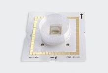
As organoid research continues to grow and evolve, the limitations of traditional 2D planar microelectrode arrays (MEA ) have given rise to the need for more advanced technologies. For researchers looking to record electrophysiological data from inside an intact organoid, Mesh MEA from Multi Channel Systems, an affiliate of Harvard Bioscience, offers significant advantages over its 2D counterparts in recording and analyzing the complex electrical activities of 3D organoids.
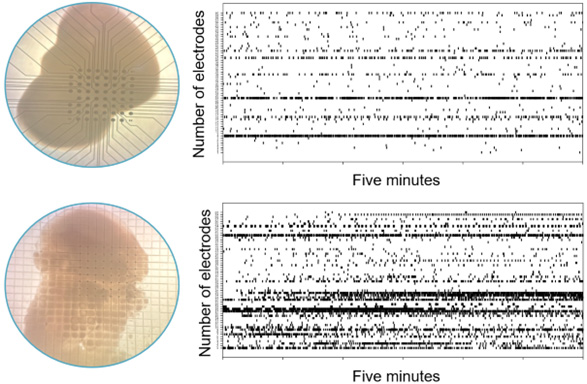
Our 2D planar MEA chips are the ideal choice for cell cultures, stem cells, tissue slices, and many other cultures, but an organoid cultured on a 2D MEA chip will collapse and flatten, which may compromise the physiological responses of the organoid and thus impact the validity of the data. Mesh MEA allows the organoid to grow around a layer of mesh, allowing it to maintain its shape while electrophysiological data is recorded from inside the organoid.

Structural Integrity and Data Quality
One of the primary advantages of Mesh MEA lies in its ability to maintain the structural integrity of organoids. Traditional 2D MEAs require the organoid to be transferred from its culture environment to the MEA platform, a process that often results in structural alterations. But Mesh MEA allows for a long-term culture to be done directly in the MEA chip, preventing deformation and preserving the organoid’s morphological structure. This ensures the collection of more relevant, true-to-life electrophysiological data from intact organoids.
Experiment Duration and Long-term Measurements
The longevity of electrophysiological recordings is another area where Mesh MEA outperforms traditional 2D MEAs. Organoids placed on 2D MEAs tend to deform within a few days , which can jeopardize long-term data recordings. Mesh MEAs, with their structural integrity and integrated perfusion systems, allow for extended monitoring periods. This capability is crucial for chronic studies, enabling researchers to conduct repeated long-term measurements and gather more comprehensive data on cellular electrophysiology activities and drug effects over time.
Organoid Accessibility and Electrode Incorporation
Traditional 2D MEAs fall short in recording data from within organoids without causing structural damage. The planar nature of these systems limits the scope of measurements to the organoid surface. Mesh MEAs, however, facilitate cellular migration around the electrode-containing mesh, allowing data recordings from the interior of the organoid. This internal recording capability provides a more accurate representation of the electrophysiological activities occurring within the 3D tissue structure.
Mitigating Experimental Risks and Reducing Costs
The manipulation required to transfer organoids to traditional 2D MEAs increases the risk of experimental errors and associated costs. Mesh MEAs mitigate these risks by enabling the generation, maturation, maintenance, and measurement of organoids within a single instrument. This streamlined process not only reduces the likelihood of costly mistakes but also spreads expenditures more effectively, making it a cost-effective solution for long-term research projects.
Operational Flexibility and Future Applications
While 2D MEAs are optimized for cell cultures and tissue slices, they are limited in their application to 3D measurements. The advanced design of Mesh MEAs supports culture at the air-liquid interface and perfusion, making them adaptable to a wide range of experimental setups.
Conclusion
Mesh MEA technology represents a significant advancement in organoid electrophysiology. By addressing the limitations of traditional 2D planar MEAs, Mesh MEAs enhance data quality, extend recording durations, and reduce experimental risks and costs. Their ability to facilitate internal recordings within 3D structures without compromising structural integrity makes them an invaluable tool for researchers. As the field progresses, Mesh MEAs are poised to play a crucial role in developing more sophisticated 3D models for studying human neurodevelopment and neurological disorders.
Incorporating Mesh MEA technology into organoid research not only improves the accuracy and relevance of electrophysiological data but also paves the way for new discoveries in basic research, drug discovery, precision medicine, and toxicology.
